Assessment Tool

  • General assessment
  • Foot inspection
  • Testing for loss of protective sensation
  • Testing for Peripheral artery disease

  • General assessment of the patient includes planning the physical/lab investigations for the patient including:

  • Their status of diabetes control (HbA1c)
  • Presence of comorbidities like hypertension / cardiovascular disorders
  • Family history of diabetes
  • Personal history of smoking
  • Nutritional assessment
  • Assessment of patient's mobility
  • History of previous foot ulceration and/ or surgery
  • Pain scoring

  • Diabetic Foot Examination

    SENSORY EXAMINATION VASCULAR EXAMINATION DEFORMITY ULCER EXAMINATION
    Vibratory perception: 128 Hz tuning fork or electronic tuning fork Pedal pulses: dorsalis pedis, posterior tibial, perforating peroneal Bunions, hammertoes, bone spurs, plantarflexed metatarsals, pes cavus foot type Area, toe, metatarsal forefoot, lateral, medial
    Achilles reflex Erythema or cyanosis Hallux limitus, Achilles/ gastro equinus, overpronation Ischemic or neuropathic or mixed
    Monofilament test 10 point touch [42] Intermittent claudication score Rocker bottom appearance Small <10 cm, moderate 11–40 cm, severe >40 cm
    Vibration perception threshold (VPT) Temperature comparison between fee Prior amputation Cool with absent pulses
    Temperature sensation Dry skin and fissuring Gait evaluation Depth; probe test
    Pain sensation Vascular Doppler ultrasonography Foot drop, atrophy, necrobiosis lipoidica diabeticorum Healing or nonhealing (inflammatory granulating epithelialization)


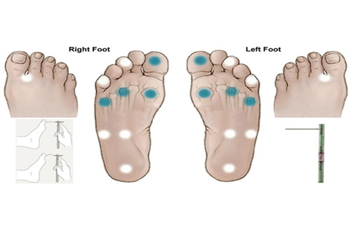
    Figure 6: Demonstration of sensation test

    Testing for Peripheral Arterial Disease
    Parameters to examine in order to detect PAD:

  • Palpation of peripheral pulses:
  •         Femoral
            Popliteal
            Posterior tibial (PT)
            Dorsalis pedis (DP)
  • Ankle Brachial Pressure Index (ABPI)
  • Toe-Brachial Index (TBI)
  • Temperature difference between the feet
  • Transcutaneous oxygen measurement (if available)

  • Doppler and Ultrasound (Duplex) imaging: a non-invasive method that visualizes the artery with sound waves and measures the blood flow in an artery to indicate the presence of a blockage.
  • Computed Tomographic Angiography (CT): a non-invasive test that can show the arteries in your abdomen, pelvis and legs. This test is particularly useful in patients with pacemakers or stents.
  • Magnetic Resonance Angiography (MRA): a non-invasive test that gives information similar to that of a CT without using X-rays.
  • Angiography: During an angiogram, also called an arteriogram, a contrast agent is injected into the artery and X-rays are taken to show blood flow, arteries in the legs and to pinpoint any blockages that may be present.

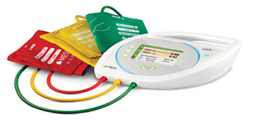
  • Figure 7: Instrument to measure ankle-brachial pressure

    Ankle-brachial pressure index

  • The ankle-brachial pressure index (ABPI) is a non-invasive method of assessing the extent of chronic peripheral arterial disease in the lower limbs.
  • It is a ratio composed of the blood pressure in the brachial artery and the pressures in the foot arteries (dorsalis pedis and the posterior tibial artery).
  • Ischemia Grade ABPI
    0 ≥ 0.80
    1 0.6-0.79
    2 0.4-0.59
    3 ≤ 0.39
    Table 1: The grading of ischemia as per the ankle-brachial pressure index.

    Measuring the Brachial Pressure

  • The sphygmomanometer cuff is placed over the left arm proximal to the brachial artery.
  • The doppler probe is positioned on the brachial artery at an angle of 45°.
  • The cuff is inflated to 20-30 mmHg above the pressure at which Doppler pulse is no longer heard.
  • Then the cuff is deflated slowly and the pressure at which the Doppler is first able to detect a pulse is noted.
  • Measuring the Ankle Pressure

  • A sphygmomanometer should be placed on the left ankle and the Doppler probe over the posterior tibial artery.
  • The procedure to measure pressure is similar to brachial artery: the cuff should be inflated and deflated slowly to detect the pressure at which blood returns to the posterior tibial artery.
  • Repeat this procedure (keeping the sphygmomanometer in the same place) at the dorsalis pedis artery of the left foot.
  • The highest of the two pressures obtained from the posterior tibial artery (PTA) and dorsalis pedis (DP) is used the ratio of the left ABPI.
  • The same process is to be repeated at the right ankle to calculate the ratio for the right ABPI.
  • The ABPI should be calculated and mention the need to document in the patient's notes.
  • Copyright © 2020 Centaur Pharmaceuticals Pvt Ltd.